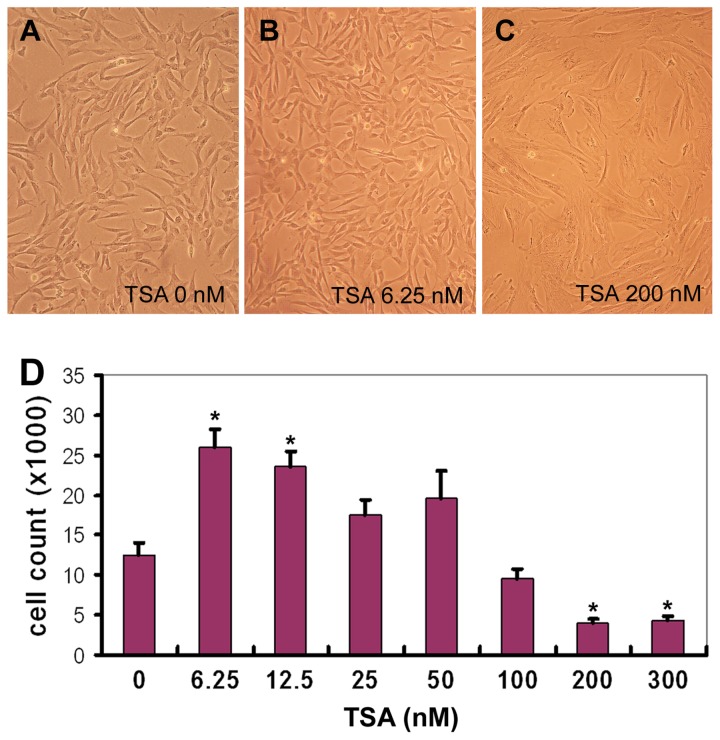
Figure 2. TSA treatment of hMSCs.
Human MSCs were grown in the presence of different concentrations of TSA (0~300 nM) for 3 days and photographed under microscope. Reprehensive images were shown (A-C). Then the cells were detached and counted. Triplet wells were used for each experiment (D, n=4, * P<0.01).

