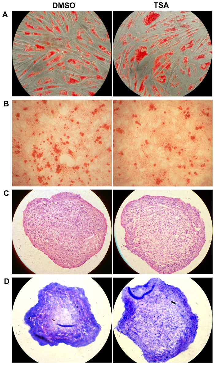
Figure 4. Differentiation of hMSCs.
Passage (P)1 hMSCs were grown in the presence of TSA (at 6.25 nM) or vehicle DMSO to passage 6. Then the cells were incubated in adipogenic (A), osteogenic (B) and chondrogenic (C and D) induction media, respectively. MSCs differentiated into adipocytes (A, with oil red staining), osteoblasts (B, with Alizarin Red S staining) and chondrocytes (cultured in micromass and sectioned: C, with H&E staining; D, with toluidine blue staining showing pink extracellular matrix proteoglycans). The experiments were repeated three times with similar results and representative results from one experiment were shown.

