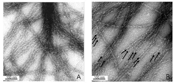
Figure 5.
S1 decoration of actin within a fesselin-actin bundle showing that actin filaments in a bundle have the same polarity. 2μM of G-actin was mixed with 0.04μM of fesselin in 40 mM KCl, 1mM MgCl2, 5mM potassium phosphate buffer, pH 7.0. After incubation in the tube for 2-3 min the sample was applied to the grid. The complex was washed with 1-2 drops of 1μM of S1 in the same buffer. Panels A and B are different fields of the same grid. Arrows on panel B show the polarity.

