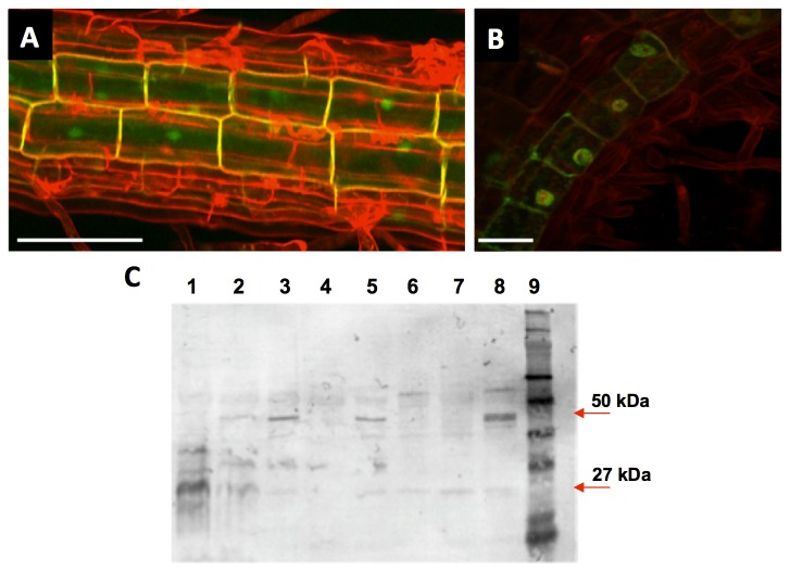
Figure 6. Subcellular location of SAUR76-protein revealed by protein-GFP fusions.
A) Projected Z-stack image of C-terminal GFP fused to the SAUR76 protein. Cell walls were counterstained with propidium iodide. B) Co-localisation of C-terminal GFP fused to the SAUR76 protein and DRAQ5, a red fluorescent nucleus stain. Scale bars are 50 and 25μm in A and B respectively. C) Western blot analysis using an anti-GFP antibody on proteins from plants bearing free GFP (lane 1) and from several independent lines containing the C-terminal protein-GFP construct (lane 2-8). Lane 9 contains the molecular marker.

