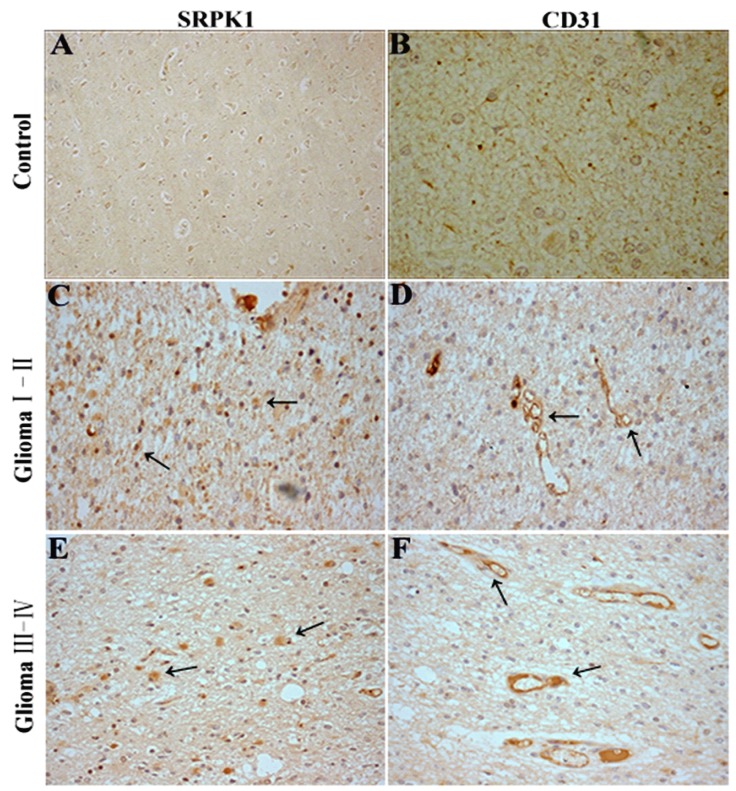
Fig 6.
SRPK1 and CD31 immunohistochemistry(IHC) in glioma tissues. (A, B) SRPK1 and CD31 IHC staining in normal brain tissues around the excision of tumor tissue. (C, D) IHC staining for SRPK1 and CD31 staining in glioma I-II tissues. (E, F) IHC staining for SRPK1 and CD31 staining in glioma III-IV tissues. All photomicrographs were taken at 200× magnification; haematoxylin counterstain. Arrows indicate the positive expression areas.

