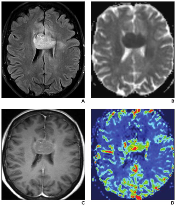
Fig. 4. 52-year-old immunocompetent man with primary corpus callosum CNS lymphoma.
A–D, Brain MRI includes transverse T2-weighted FLAIR image (A), apparent diffusion coefficient (ADC) map (B), transverse contrast-enhanced T1-weighted image (C) (0.1 mmol of gadobutrol), and relative cerebral blood volume (rCBV) map (D). Homogeneous corpus callosum mass is hypointense on ADC map, consistent with restricted diffusion. Mild contrast uptake and increased signal intensity on color rCBV map indicate hyperperfusion.

