
Figure 4.
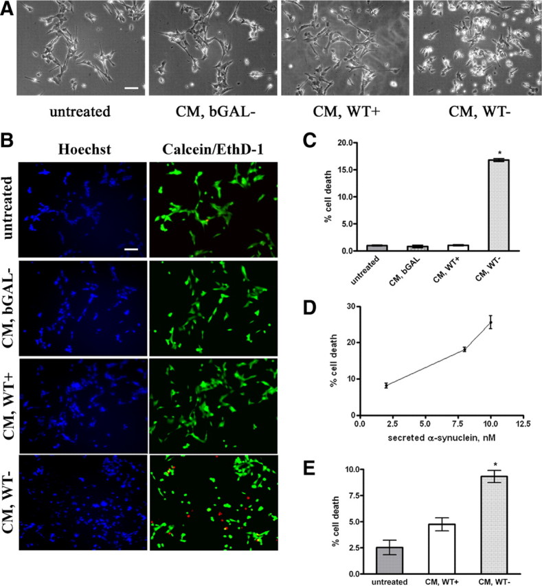
Secreted α-synuclein induces cell death to differentiated SH-SY5Y cells. A, α-Synuclein-expressing and uninduced CTL cells, as well as bGAL-expressing cells, were cultured in 2% FBS for 48 h. CM was collected and applied to differentiated SH-SY5Y cells for 24 h. Representative phase micrographs of the recipient cells are shown. Scale bar, 50 μm. B, CM was prepared and used to treat recipient SH-SY5Y cells as in A. After 24 h, the recipient cells were stained with EthD-1 (red) and Hoechst (blue). Scale bar, 50 μm. C, Percentage cell death was determined by counting the percentage ratio of EthD-1-positive cells versus the Hoechst-positive cells. Quantitative analysis demonstrated a statistically significant increase in cell death when recipient cells were treated with CM from α-synuclein-expressing cells (n = 4; mean ± SD, one-way ANOVA test followed by Tukey's test, *p < 0.001). D, CM was collected from α-synuclein-expressing cells as in A and applied to differentiated SH-SY5Y cells for 24 h. Cell death was quantified by EthD-1/Hoechst staining. The amount of secreted α-synuclein present in the CM was determined by Western immunoblotting and recombinant α-synuclein as standard. The average of three experiments is shown. E, CM from α-synuclein-expressing or uninduced CTL cells were applied to cycling SH-SY5Y cells for 24 h, and the recipient cells were stained with EthD-1 and Hoechst. Quantitative analysis showed a reduction in cell viability from α-synuclein-rich CM (n = 4; mean ± SD, independent t test, *p < 0.01).