
Figure 7.
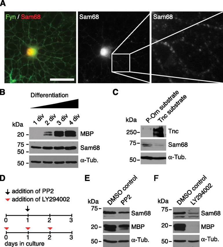
Sam68 expression increases with oligodendrocyte differentiation and is repressed through Tnc and inhibitors of SFK and PI3K. A, A Fyn-expressing oligodendrocyte coexpresses Sam68 protein. Most Sam68 immunoreactivity is restricted to the nucleus, although cytosolic Sam68 is also detectable. B, Immunoblotting of differentiating OPC cultures lysed after various cultivation periods showing that Sam68 at all stages of oligodendrocyte differentiation. Note, however, that Sam68 levels increase with differentiation (defined by increasing MBP levels). C, Immunoblottings of oligodendrocytes cultured on P-Orn or Tnc substrate for 24 h reveal that cultivation on Tnc reduces Sam68 expression levels. D, Schematic drawing of the cultivation protocol used for addition of PP2 or LY294002. E, F, Immunoblotting shows that oligodendrocytes display lower Sam68 expression levels in the presence of the SFK inhibitor PP2 or the PI3K inhibitor LY294002. Tubulin served as a loading control. Also note that the Sam68 antibody yielded one (B) or two bands (C, E, F), depending on which Sam68 antibody was used (see Materials and Methods). Scale bar, 50 μm.