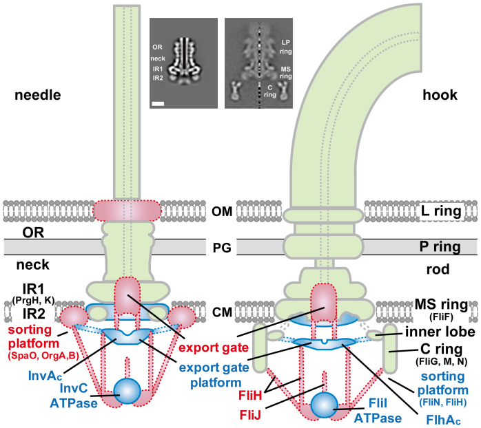
Figure 1. Schematic representation of the structures of Salmonella injectisome (NC, left) and flagellar hook basal body (FBB, right).
Different colors indicate structures of different categories: pale green, structures of isolated complexes; blue, additional structures present in the in situ complexes; red, putative structures not yet visible in the in situ complexes. Insets are the axial sections of isolated NC10 and FBB obtained by cryoEM image analysis. OR: outer membrane ring, IR: inner membrane ring, OM: outer membrane, PG: peptidoglycan layer, CM: cytoplasmic membrane.

