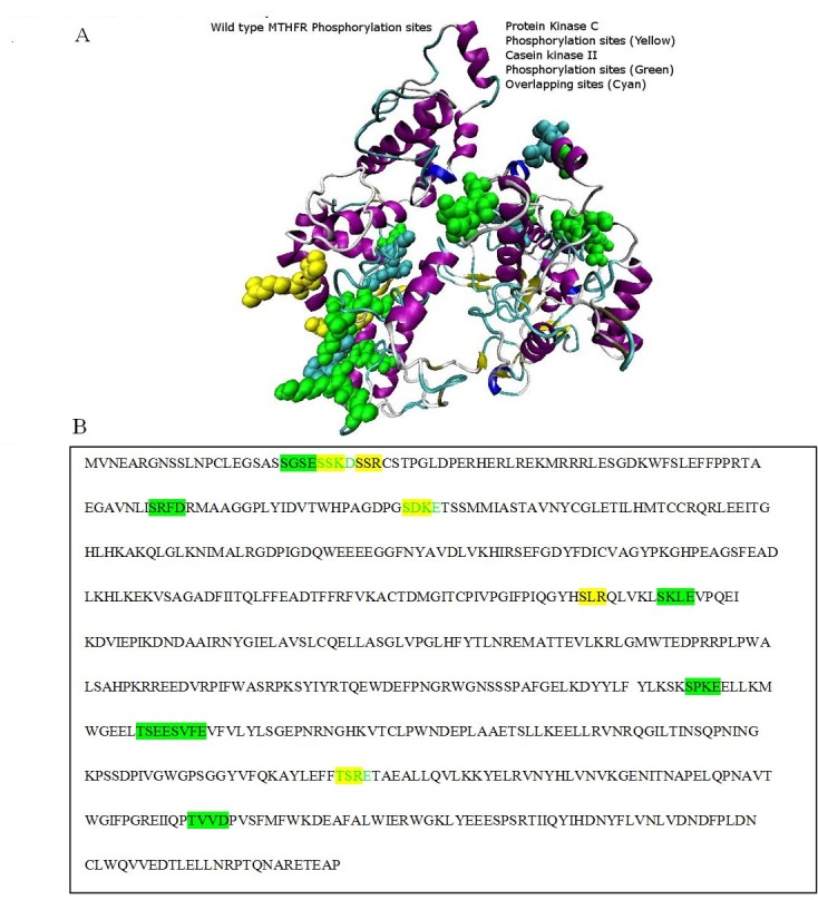
Figure 4.

Phosphorylation binding sites: A) Protein Kinase C phosphorylation sites are colored in yellow, Casein Kinase II phosphorylation sites are colored in green, while the overlapping phosphorylation sites of the both enzymes are colored in cyan as show in the figure A; B) Protein Kinase C phosphorylation sequence is colored in yellow, Casein Kinase II phosphorylation sequence is colored in green, while the green colored sequence highlighted with yellow background is the overlapping sequence of both the enzymes.
