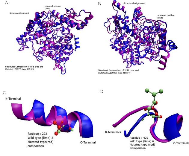
Figure 7.
A) Structural alignment of Normal (Blue) and Mutated (Purple) amino acids of MTHFR. Residue number 222 of both types is shown (CPK view). Normal amino acid Alanine is shown in the lime color while mutated amino acid Valine is shown in the red color; B) Structural alignment of the normal and mutated amino acids of MTHFR. Glutamic Acid is shown in the lime color which is replaced by Alanine in the mutated structure; C) Close look of A222V mutated structure. Blue colored helix shows the normal structure while purple colored helix represents the mutated structure; D) Close look of E429A mutated structure. Blue colored helix shows the normal structure while purple colored helix represents the mutated structure.

