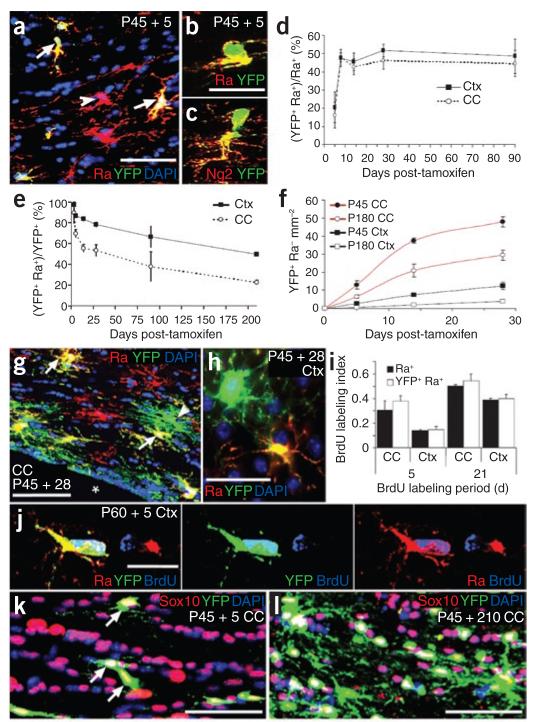
Figure 3.
PDGFRA+ adult OLPs generate differentiated oligodendrocyte lineage cells. (a–c) Sections of P45 tamoxifen-induced Pdgfra-creERT2/Rosa26-YFP mice were immunolabeled for YFP and PDGFRA or NG2 at P45 + 5 (arrows in a indicate YFP+ PDGFRA+ OLPs (yellow), arrowhead indicates a PDGFRA+ YFP− OLP (red)). (d) The fraction of PDGFRA+ cells that coexpressed YFP increased between P45 + 5 and P45 + 8, and then remained constant until at least P45 + 90, indicating that Cre recombination continues for 1–4 d after the final dose of tamoxifen. With our protocol, ~45% of OLPs labeled for YFP after P45 + 8. The fraction of YFP+ cells that coexpressed PDGFRA and NG2 was high at P45 + 3, but declined with time post-tamoxifen (e), as increasing numbers of YFP+, PDGFRA− cells appeared (f–h) (arrows in g indicate YFP+ PDGFRA+ OLPs, yellow; arrowhead indicates a YFP+ PDGFRA− cell, green). This suggests that PDGFRA+ OLPs differentiate and downregulate PDGFRA. (h) YFP+ PDGFRA+ and YFP+ PDGFRA− cells in the cortex. The rate of addition of PDGFRA− (differentiated) cells was higher at P45 than at P180 and was higher in corpus callosum than in cortex at either age (f). (i,j) The subpopulation of OLPs that were labeled for YFP was not biased toward either dividing or nondividing OLPs, as the BrdU labeling index of YFP+ PDGFRA+ cells matched that of the PDGFRA+ population overall. Between P45 + 5 and P45 + 210, all YFP-labeled cells were SOX10+ oligodendrocyte lineage cells, both in the corpus callosum (k,l) and motor cortex (data not shown). Scale bars represent 35 μm in a, g, k and l, and 10 μm in b, c, h and j.

