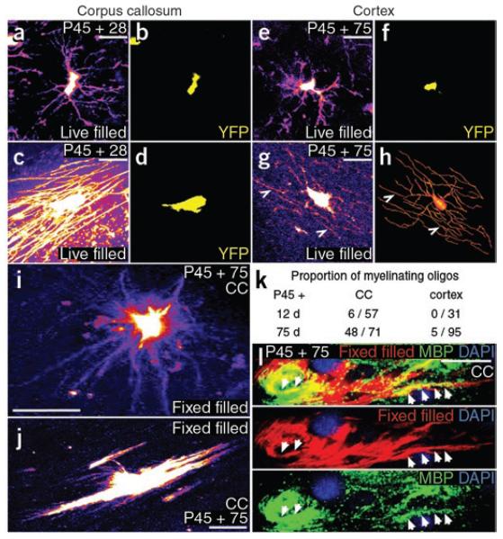
Figure 5.
Adult OLPs generate myelinating oligodendrocytes. To reveal full cell morphology, we used YFP in live tissue slices to target cells for Alexa Fluor dye injection via glass patch pipettes. (a–d) Cells with the distinctive morphology of myelinating oligodendrocytes, with up to 50 rod-like myelin internodes, were identified in the corpus callosum (c,d), as well as presumptive OLPs with slender, multiply branching processes (a,b). (e,f) In the cortical gray matter, the large majority of live dye-filled cells resembled OLPs. (g,h) A few filled cells in cortical layers 5/6 did possess some myelin-like structures (arrowheads in g); a digital reconstruction gives a clearer impression of whole cell morphology (h). (i,j) To quantify the number of cells with myelinating morphology in the corpus callosum, we filled YFP+ cells in fixed tissue slices with DiI. This allowed larger numbers of cells to be examined, but the DiI did not perfuse the cells as completely as did Alexa dye in live cells. Nevertheless, we could distinguish cells with progenitor morphology (i) versus myelinating morphology (j). (k) The proportion of myelinating cells increased with time post-tamoxifen in the corpus callosum; at P45 + 75 roughly 70% of YFP+ cells had myelinating morphology. (l) We immunolabeled fix-filled cells with antibody to MBP and showed that dye-filled processes contained MBP, indicating that they were indeed myelin internodes. Scale bars represent 10 μm.

