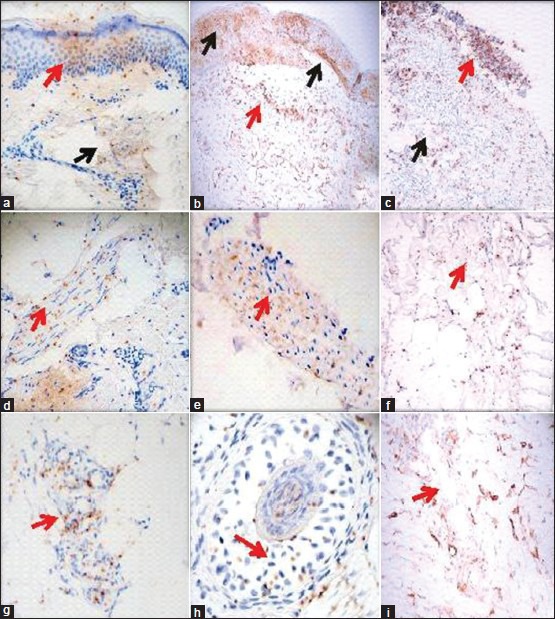
Figure 1.

Positive staining of S6-pS240 immunohistochemistry (IHC) in multiple autoimmune blistering diseases. All IHC stains were performed utilizing the monoclonal mouse antihuman ribosomal protein S6-pS240. (a) A representative case of pemphigus foliaceus (PF), showing reactivity in a dermal eccrine sweat duct (brown staining, black arrow) and in the epidermal stratum granulosum and corneal layer (brown staining; red arrow). (b) A representative case of PV, demonstrating reactivity in the epidermis in proximity to a pathogenic blister, as well as on the blister roof (brown staining; black arrows) and in a mesenchymal/endothelial cell junction-like structure in the dermis (brown staining; red arrow). (c) A BP case, displaying positivity in a subepidermal blister area (brown staining; red arrow) and in a dermal mesenchymal/endothelial cell junction-like structure (brown staining; black arrow). (d and e) Two different cases of El Bagre-endemic pemphigus foliaceus (EPF), demonstrating reactivity in dermal piloerector muscles (brown staining; red arrows). (f) A DH case, highlighting positivity in a dermal endothelial-mesenchymal cell junction-like structure (brown staining; red arrow). (g and h) Two cases of El Bagre EPF, demonstrating reactivity within a dermal eccrine gland coil and a hair follicle, respectively (brown staining; red arrows). (i) A case of BP, displaying positivity to a dermal mesenchymal-endothelial cell junction-like structure (brown staining; red arrow)
