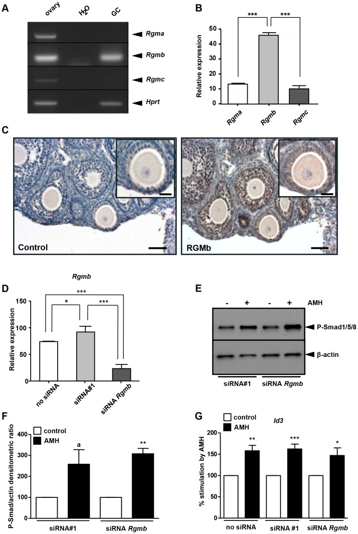
Figure 6. RGMb is not essential for AMH signalling in granulosa cells.
Rgma, b and c expression was analysed by RT-PCR (A) or real time PCR (B, n = 6). (A) RT-PCR showed that all three Rgm were expressed in the mouse immature ovary (left lane) while Rgmb seemed predominantly expressed in GCs (right lane) (A). Real-time PCR confirmed that Rgmb was more expressed than Rgma and Rgmc in GCs (B). Data were analyzed using one-way ANOVA followed by Tukey test for all-pair comparisons. Mouse immature ovary was subjected to immunohistochemistry using an RGMb antibody (C). The left panel was the control without the primary antibody. The right panel shows that RGMb is expressed in the cytoplasm of granulosa cells (scale bar = 50 µm; insert, scale bar = 20 µm). siRNA transfection targeting Rgmb was performed when cells were 50% to 80% confluent (D–G). GCs were exposed (▪) or not (□) to 8 nM AMH. Real-time PCR was used to quantify the decrease in Rgmb expression. Rgmb expression dropped about 75% in GC transfected with the siRNA targeting Rgmb when compared to a control siRNA (D, n = 6). Western blot with a phospho-Smad1/5/8 antibody (E, n = 4) and real-time PCR on Id3 gene (G, n = 6) showed that the knowkdown of Rgmb does not affect AMH signalling pathway. Western blots were quantified and normalized from 4 experiments (F, n = 4). Data were analyzed using paired t-test. * p<0.05, ** p<0.01, *** p<0.001, a: p = 0.074. AMH response was not significantly different between control siRNA and Rgmb siRNA transfected GCs.

