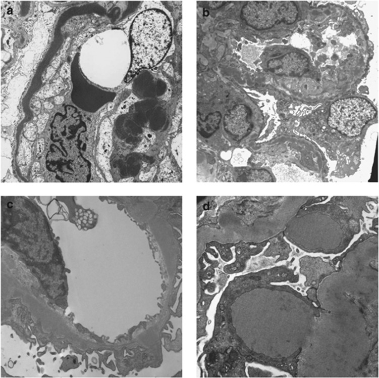
Figure 2.
Electron microscopy in C3 glomerulopathy. (a) Dense deposit disease showing very electron-dense osmiophilic deposit occupying most of the glomerular basement membrane. (b) A case of dense deposit disease in which the highly osmiophilic deposit is only seen in segments of the glomerular basement membrane. (c) A case of C3 glomerulopathy in which there is electron-dense material that is expanding the basement membrane. The material is less electron dense and less well defined than in dense deposit disease. (d) A case of C3 glomerulopathy showing two large subepithelial hump-shaped deposits.

