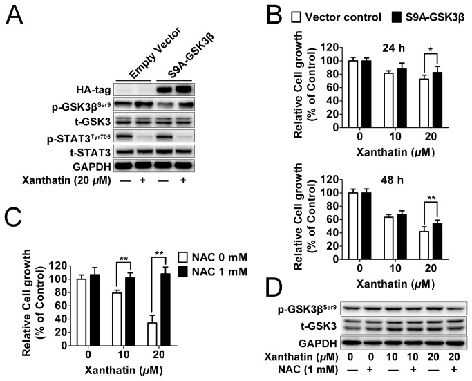
Figure 5. GSK3β is essential for the anticancer effect of xanthatin in A549 cells.
(A) A549 cells seeded in 6 well plate were transfected with HA-tagged constitutively active (S9A)-GSK3β or empty vector (2 μg plasmid DNA per well). After for 48 h post transfections, cells were treated with or without 20 μM xanthatin for 6 h, then were subjected to Western blot for measuring protein levels of HA tag, phosphor-STAT3 (Tyr705) and STAT3 respectively. (B) A549 cells seeded in 96 well plate were transfected with HA-tagged constitutively active (S9A)-GSK3β or empty vector (0.1 μg plasmid DNA per well). After for 48 h post transfections, cells were treated with 10 or 20 μM xanthatin for 24 h and 48 h respectively, then were subjected to cell proliferation assay. For indicated comparisons, *P<0.05, **P<0.01. (C) A549 cells were treated with 10 or 20 μM xanthatin in presence or absence of 1 mM NAC for 48 h, then were subjected to cell proliferation assay. (D) A549 cells were treated with 10 or 20 μM xanthatin in presence or absence of 1 mM NAC for 6 h, then were subjected to Western blot for measuring protein levels of phosphor-GSK3β (Ser9) and GSK-3 respectively. The data shown are represented as the mean ± SD. For indicated comparisons, *P<0.05, **P<0.01.

