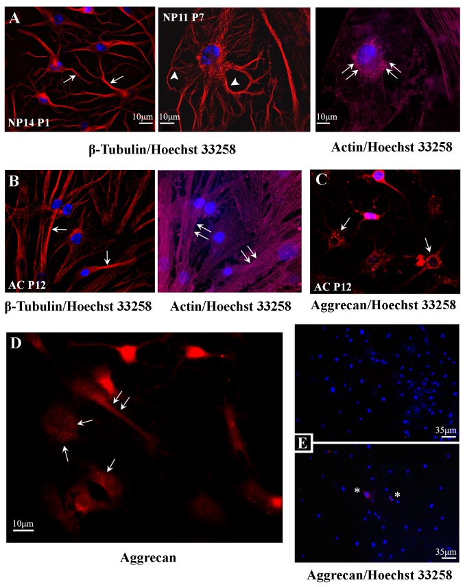
Figure 2. Cytoskeletal and chondrocyte phenotypic markers in cultured NP cells.
NP cell cultures were fixed, immunostained and imaged with deconvolution microscopy (A) β-tubulin immunostaining (red) reveals elaborate microtubule cytoskeletons in NP cells (arrows), especially in the rare large notochordal-like cells (arrowheads), while phalloidin staining (magenta) shows that F-actin is organized in focal adhesions under the cell body (double arrows); nuclei are stained blue with Hoechst. (B) In contrast, AC cells exhibit polarized cell bodies, lesser microtubule branching (arrows), and F-actin forms intense stress fibers (double arrows). Staining with the BC-3 antibody (red) that recognizes the neoepitope on human aggrecan that is created after cleavage within the sequence TEGE373-374ARGS by aggrecanases activity, decorated the cytoplasm of both (C) AC cells and (D) NP cells. In both cell types the staining pattern was consistent with a vesicular distribution (arrows). (E) Aggrecan staining was not detected when chondroitinase was omitted (upper panel) or when the reaction was allowed to proceed at 4° C (asterisk indicate cells at mitosis) (NP6 P10, C7). NP tissue indicate fresh tissue sample, NP numbers indicate patient sample, and P the culture passage; all cells were visualized with a 63x oil immersion lens.

