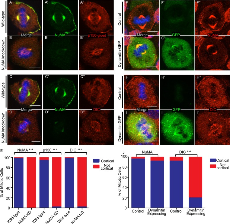
FIGURE 1:
NuMA recruits dynein/dynactin to the cell cortex of keratinocytes. (A–D) Immunofluorescence analysis of endogenous NuMA, p150glued, and DIC localization in wild-type and NuMA-knockdown mouse keratinocytes, as indicated. (E) Quantitation of cells with cortical NuMA, p150-glued, and DIC localization. n = 50 cells for each, p < 0.0001 for each. (F–I) Immunofluorescence analysis of endogenous NuMA and DIC localization in untransfected and dynamitin-GFP–transfected cells. (J) Quantitation of cells with cortical NuMA and DIC localization. n = 25 cells for each, p = 1 for NuMA, p < 0.0001 for DIC. Scale bars, 10 μm.

