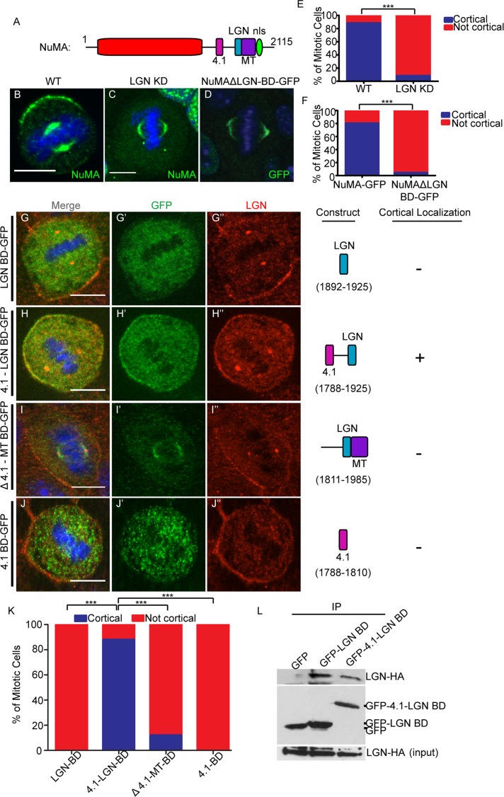
FIGURE 2:
LGN binding is necessary but not sufficient for cortical NuMA recruitment, which may require association with 4.1. (A) The characterized binding regions within the NuMA protein. (B, C) Immunofluorescence analysis of endogenous NuMA localization in wild-type (WT) and LGN-knockdown keratinocytes. (D) Localization of GFP-tagged NuMA lacking the LGN-binding domain (ΔLGN BD-GFP) in wild-type cells. (E) Quantitation of NuMA cortical localization in WT and LGN-knockdown cells. n = 50 cells, p < 0.001. (F) Quantitation of cortical NuMA-GFP and NuMAΔLGN-BD-GFP localization. n = 25 cells, p < 0.0001. (G–J) Various truncation constructs of NuMA (see Construct column) tagged to GFP were transfected into wild-type cells. The amino acids spanned in each construct are specified in the Construct column. Cells were stained for endogenous LGN, and subsequent immunofluorescence analysis was performed to compare localization of these constructs with respect to cortical LGN. The Cortical column indicates whether cortical localization was detected for each construct (+, presence in; –, absence from cortex). (K) Quantitation of cortical localization of NuMA deletion constructs, as indicated. n = 25 cells for each, p < 0.0001 when comparing the 4.1-LGN BD to either the LGN BD or Δ4.1-MT BD. Scale bars, 10 μm. (L) Immunoprecipitation of GFP-tagged LGN-BD– and 4.1-LGN BD–transfected keratinocytes. Lysates were probed with anti-HA antibodies to detect associated LGN-HA. Middle blot, amounts of GFP fusion proteins in the immunoprecipitates; bottom, levels of LGN-HA in the lysates.

