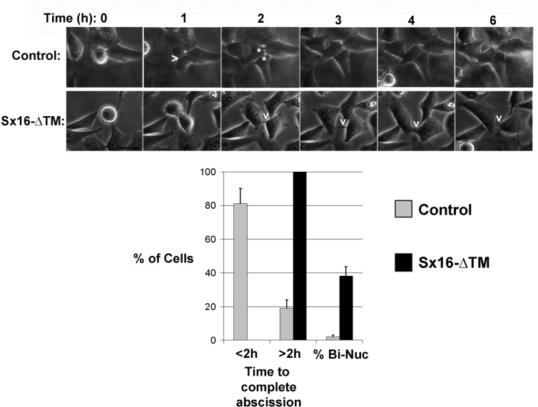
FIGURE 2:
Sx16 is involved in abscission. Images from a typical time course of HeLa cell division, revealing delayed abscission in Sx16-ΔTM–expressing cells (bottom) compared with control cells (top). In control cells, a furrow is indicated by >, and cytokinesis is complete ∼1 h thereafter (two daughter cells indicated by asterisks in the next image). In Sx16-ΔTM–expressing cells, a long-lived midbody is indicated by the v; in the experiment shown, the cells remained connected for >4 h from the first clear appearance of the midbody (shown in the 2-h image). Data from >25 cells from three or more experiments in each group were binned according to the time taken to complete abscission after the start of furrowing into two groups, those taking <2 h, or those taking >2 h. The frequency of binucleate cells in this subgroup is also presented. Cells expressing Sx16-ΔTM consistently took longer to complete abscission, or failed abscission at a significantly greater rate, than their control counterparts.

