FIGURE 1:
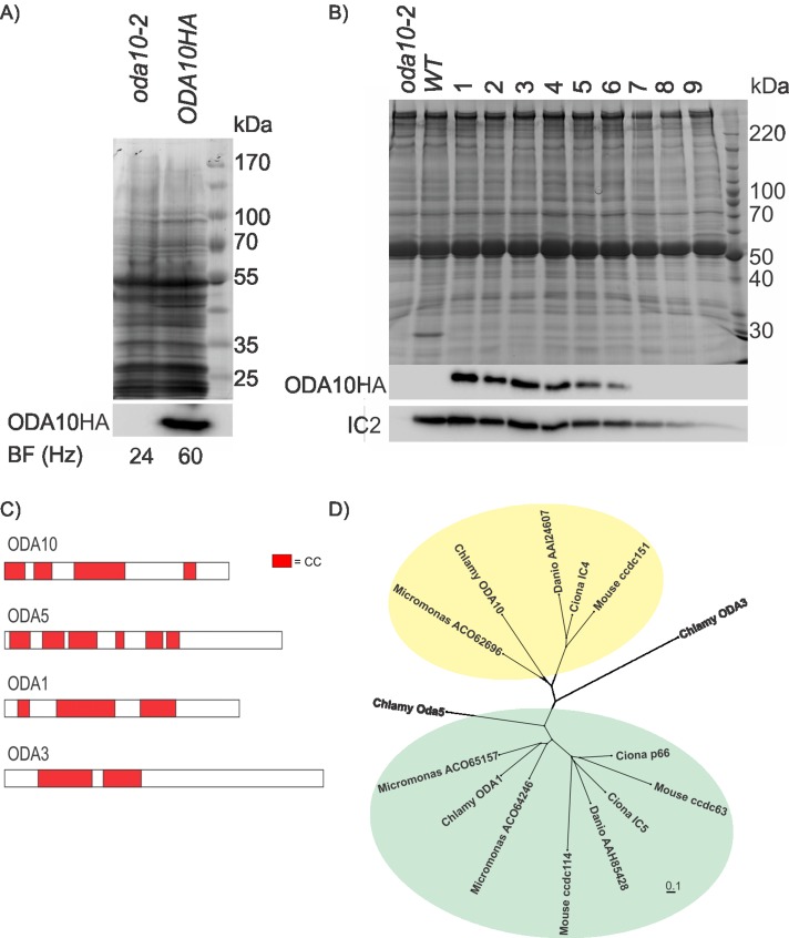
ODA10p is a conserved coiled-coil protein. (A) A Coomassie-stained gel (top) and Western blot (bottom), probed with an antibody against HA epitope, of equally loaded (by cell number) whole-cell samples of oda10-2,ODA10HA and oda10-2 cells show expression of ODA10HAp in a strain with rescued beat frequency. (B) A Coomassie-stained gel (top) and western blot (bottom), probed with antibodies against HA epitope and IC2, of flagellar samples of mutant (oda10-2), wild type (137c), and rescued transformants (oda10-2,ODA10HA 1-9) show restored assembly of outer dynein arms to wild-type levels in transformant cells expressing the highest levels of ODA10HAp. (C) The distribution of predicted coiled-coil domains of ODA1p, ODA3p, ODA5p, and ODA10p. (D) All homologues of ODA10p and of three Chlamydomonas ODA10p paralogues (ODA5p, ODA1p [ = DC2], ODA3p [ = DC1]) in an alga (Micromonas), a basal chordate (Ciona), a fish (Danio), and a mammal (Mus) were aligned, and an unrooted tree was generated to display relative sequence similarities. Green background highlights homologues of ODA1p; yellow background highlights homologues of ODA10p. Protein sequences used in the alignments that are not given in the figure were Q8CDV6 (ccdc63), AAI60348 (ccdc114), AAH57069 (ccdc151), NP_001027646 (Ciona p66), NP_001072015 (Ciona IC4), NP_001071888 (Ciona IC5), AAC49732 (ODA3), AAK72125 (ODA1), AAS10183 (ODA5), and AGW18228 (ODA10).

