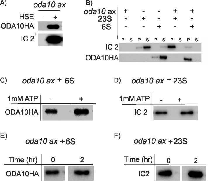
FIGURE 4:
ODA10p and outer arm dynein stably assemble onto oda10 axonemes independently in vitro. Western blots probed with anti-IC2 and anti-HA detect proteins bound in vitro to oda10 axonemes. (A) oda10 axonemes were incubated with buffer alone (–) or a dialyzed HSE of wild-type axonemes from ODA10HA cells (+) for 1 h, and pellet fractions were examined. Both IC2 and ODA10HAp are present in the pellet. (B) oda10 axonemes were incubated on ice for 1 h with separate sucrose gradient fractions of outer row dyneins (∼23 S) or ODA10HAp (∼6 S) or buffer alone. Both pellet (P) and supernatant (S) were analyzed. (C, D) The binding experiment in B was repeated in presence of 1 mM ATP, and pellets were probed for ODA10HA or IC2. ODA10HA and outer dynein arms pellet with axonemes, regardless of presence of ATP. (E, F) oda10 axonemes previously incubated with either 6S (E) or 23S (F) fractions were pelleted, and half of each sample was resuspended in fresh buffer, incubated on ice for 2 h, and pelleted again. Equal amounts of ODA10HAp (E) and IC2 (F) are associated with oda10 axonemes before (0) and after (2) incubation.

