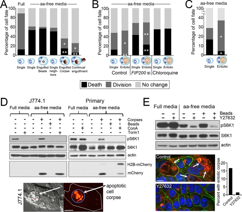
FIGURE 1:
Cell engulfment rescues the effects of amino acid deprivation. (A) Engulfment of apoptotic corpses rescues macrophages from amino acid deprivation–induced cell death. Fates of J774.1 macrophages cultured in full and amino acid (aa)–free media in the presence or absence of apoptotic corpses, determined by time-lapse microscopy for 48 h. Single, single cells, n = 90 for full and 90 for aa-free media; engulfed beads, cells that engulfed from one to five latex beads, n = 60; single neighbors, single cells within same microscopic fields as corpse-engulfing cells, n = 168; engulfed corpse, cells with one or two corpses engulfed before start of time lapse, n = 90; continual engulfment, cells supplied with corpses that were engulfed continuously throughout the time lapse, n = 74. *p < 0.02, **p < 0.001 (when compared with single cells in aa-free media; chi-squared). Data are from at least three independent experiments. (B) Entosis rescues MCF10A cells from the effects of amino acid deprivation. Fates of MCF10A cells (single) and MCF10A cells with an entotic cell corpse (entotic) in aa-free media time lapsed for 48 h (control and FIP200 siRNA [FIP200 si]–treated cells) or 18 h (chloroquine-treated cells). Control single cells, n = 360; control entotic, n = 137; FIP200si single, n = 558; FIP200si entotic, n = 179; chloroquine single, n = 92; chloroquine entotic, n = 37. *p < 0.002, **p < 0.001 (chi-squared). Data are from at least three independent experiments. (C) Entotic MCF-7 cells (n = 192) harboring an entotic corpse are rescued from cell death and proliferation arrest compared with single control cells (n = 567) in aa-free media. p < 0.001 (chi-squared). Cells were examined for 48 h by time-lapse microscopy. Data are from at least three independent experiments. (D) mTORC1 is reactivated in aa-free media by corpse digestion in J774.1 macrophages (left blots) and primary bone marrow–derived macrophages (right blots). Western blots show restoration of phosphorylated S6-kinase threonine 389 (pS6K) by apoptotic corpse engulfment but not latex bead engulfment in macrophages cultured in aa-free media. pS6K restoration is blocked by treatment with the mTOR inhibitor Torin1 and the lysosome inhibitor ConA. Torin1 was added to cultures 1 h before cell lysis; ConA was added for the duration of the experiment. Untreated macrophages digest the corpse-specific marker H2B-mCherry into free mCherry protein, which is inhibited by ConA treatment. Images show apoptotic corpse expressing H2B-mCherry (red fluorescence, arrow) engulfed by J774.1 macrophage. Bar, 10 μm. (E) mTORC1 is reactivated by entosis. Western blots show higher levels of pS6K in MCF-7 cells cultured in aa-free media under control conditions, with 15% of cells harboring entotic corpses (quantified in graph; representative of two independent experiments), compared with entosis-inhibited conditions with Y-27632 treatment. Images show entotic cell corpses (white arrows) in control cultures, which are absent from Y-27632–treated cultures. Immunofluorescence staining for Lamp1 (red) and β-catenin (green) and 4′,6-diamidino-2-phenylindole (DAPI)–stained nuclei (blue). Bar, 10 μm.

