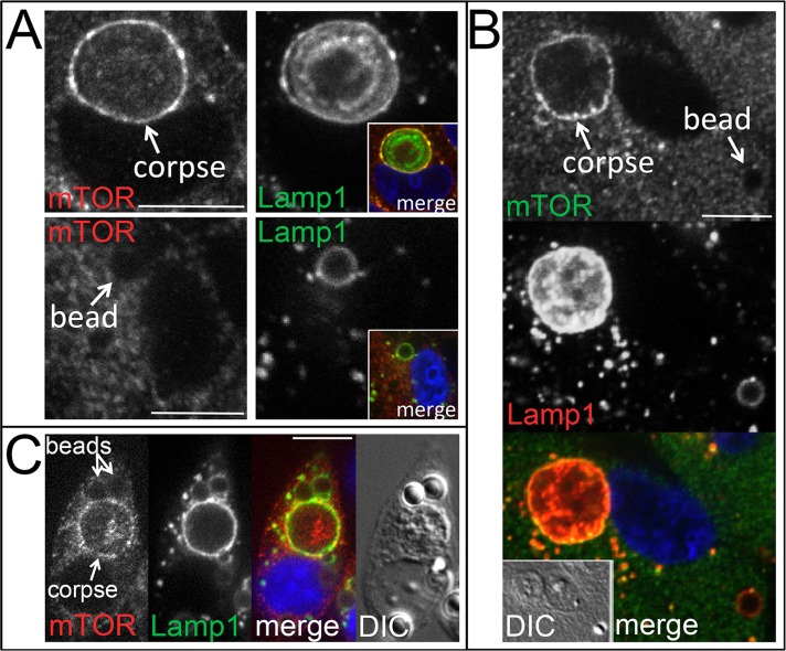
FIGURE 2:
mTOR is recruited to lysosomal vacuoles harboring engulfed cells but not latex beads. (A) Top, mTOR (left) is recruited to corpse-containing entotic vacuole (arrow) (MCF10A), where it colocalizes with Lamp1 (right); inset, colocalization of immunostained mTOR (red) and Lamp1 (green). DAPI-stained nucleus is shown in blue. Bottom, mTOR does not recruit to bead-containing vacuole (left, arrow), which is marked by Lamp1 (right) (MCF10A); inset, merged image with DAPI-stained nucleus in blue. All are confocal microscopic images. Bars, 10 μm. (B) mTOR localizes to corpse-containing entotic vacuole (labeled corpse with arrow; 70% were positive for mTOR, n = 37) but not to bead-containing lysosomal vacuole (bead, arrow; 2.3% positive for mTOR, n = 84) in the same MCF10A cell. Top and middle, confocal images of immunofluorescence for mTOR and Lamp1; bottom, merge with DAPI-stained nucleus (blue); inset, DIC. (C) mTOR localizes to apoptotic cell phagosomes (52% positive for mTOR, n = 54) but not latex bead phagosomes (1.9% positive for mTOR, n = 154) in J774.1 macrophages. Confocal microscopic images show macrophage with an engulfed apoptotic corpse and two beads, as indicated, stained for mTOR and Lamp1 by immunofluorescence. Right, merged image with DAPI-stained nucleus (blue) and DIC.

