FIGURE 1:
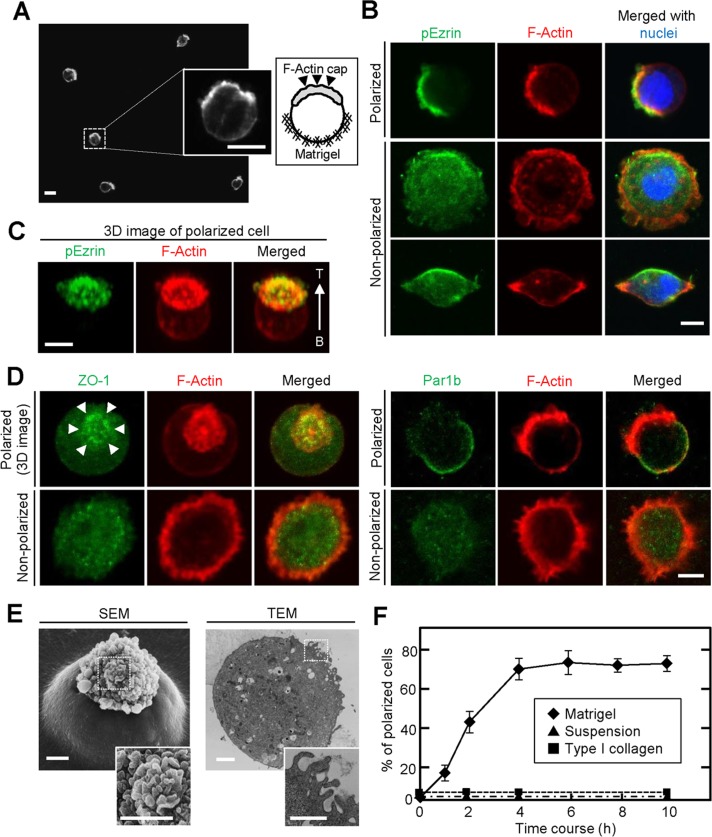
IEC6 cells show a polarized cell shape at the single-cell level in a Matrigel-dependent manner. (A) IEC6 cells cultured on Matrigel for 8 h were stained for F-actin. Left and right inserts, enlarged view and schema, respectively, of the area within the dashed box. (B) IEC6 cells were plated on Matrigel for 2 h and stained for pEzrin (green), F-actin (red), and DRAQ5 (blue). Top, a typical polarized cell; bottom, unpolarized cells. (C) Three-dimensional image of typical polarized IEC6 cells generated from an image stack. T, top; B, bottom. (D) IEC6 cells were plated on Matrigel and stained for ZO-1 (green, left) or Par1b (green, right) and F-actin (red). (E) The presence of microvillus structures on top of single IEC6 cells was observed by SEM (left) and TEM (right). Bottom, enlargement of images within the dashed boxes, indicating the apical surface. (F) IEC6 cells were suspended in growth medium (▲) or plated on Matrigel (♦) or type I collagen gel (■) for indicated time periods. Polarized cells among at least 100 cells were counted at indicated time points and expressed as the percentage of polarized cells. Results are means ± SD from three independent experiments. Scale bars, 10 μm (A), 5 μm (B–D), 2 μm (E).

