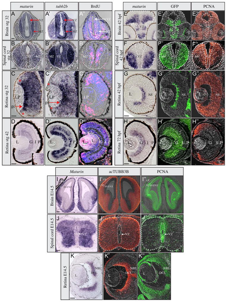
Fig. 2.
Maturin expression during neural differentiation. Expression of maturin (A–K), differentiation (A′–K′) and proliferation markers (A″–K″) in frog (A–D″), zebrafish (E–H″) and mouse (I–K″) neural tissues. Expression of maturin (A–K) and tubb2b (A′–D′) was determined using in situ hybridization. Differentiating neurons were identified by GFP immunolabeling to detect elav3l:GFP transgene expression in zebrafish (green in E′–H′) and by acTUBB3b (orange in I′–K′) immunolabeling in mouse. Proliferating neuroblasts were identified by either BrdU in Xenopus (pink in A″–D″) or PCNA immunolabeling in zebrafish and mouse (orange in E″– H″ and green in I″– K″). Nuclei were stained blue with DAPI (A″–D″). Inset in panel D is magnified view of region encompassing the dorsal CMZ. MZ, marginal zone; VZ, ventricular zone; LP, lens placode; gc, ganglion cells; *, retinal periphery; L, lens; G, ganglion cell layer; I, inner plexiform layer; P, photoreceptor layer; NE, neuroepithelium; CP, cortical plate; IZ, intermediate zone; DCL, differentiated cell layer; NBL, neuroblastic layer. Scale bars, 50 μm.

