FIGURE 3.
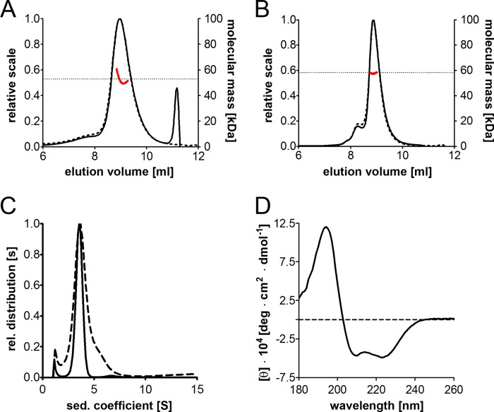
Oligomeric state and secondary structure of BAG-6686–936. A and B, SEC-MALS measurements of IMAC-purified E. coli (A) and StrepTactin-purified High Five BAG-6686–936 (B). The UV absorbance (dashed line) and the differential refractive index (solid line) were analyzed, and the resulting molecular weight affiliations (red) are indicated. C, sedimentation velocity measurements of E. coli BAG-6686–936 (dashed line) and High Five BAG-6686–936 (solid line). The main protein species has a sedimentation coefficient of 3.5 s. rel., relative. D, CD spectroscopy of BAG-6 proteins. The far UV-CD spectrum between 178 and 260 nm (at 22 °C) of E. coli BAG-6686–936 shows a mainly α-helical folding. deg, degree.

