
FIGURE 6.
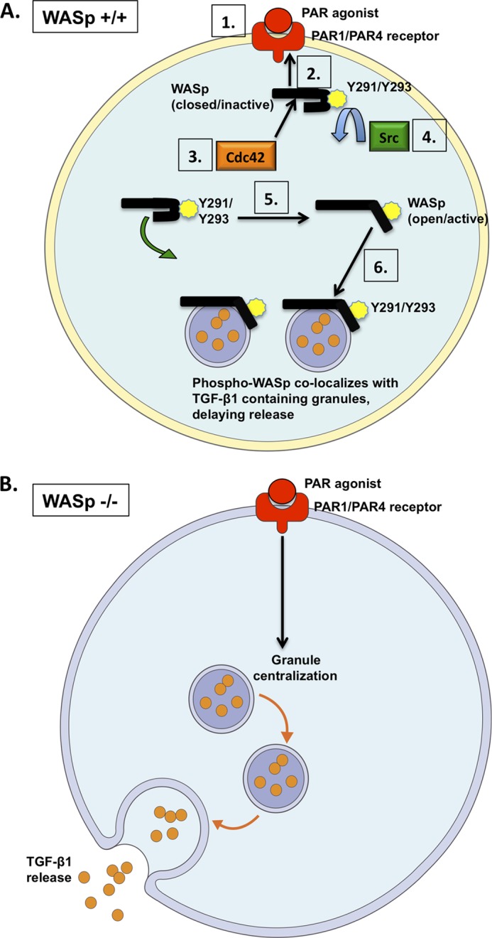
Model for WASp-mediated release of TGF-β1 in platelets. A, WASp is recruited to the PAR1/PAR4 receptor following agonist stimulation (steps 1 and 2). WASp is depicted in its closed, inactive conformation. Binding of Cdc42 to WASp (step 3) promotes the phosphorylation of WASp at Tyr-291/Tyr-293, which is mediated by Src family kinases (step 4). Phosphorylation transforms WASp into its open, active conformation (step 5). Phospho-WASp interacts with TGF-β1-containing α granules to control their spatial and temporal release (step 6). B, in the absence of WASp (or following WASp inactivation), TGF-β1-containing granules are freely trafficked within the platelet cytosol (marked by premature granule centralization and increased cytokine release).