
FIGURE 9.
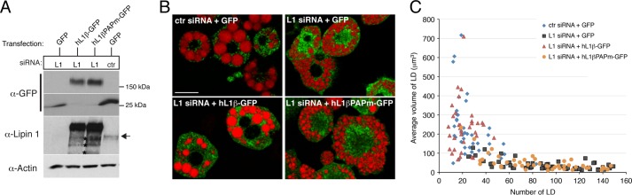
The PAP activity of lipin 1 is essential for normal lipid droplet size and number in 3T3-L1 adipocytes. A, 3T3-L1 preadipocytes were transfected with retroviral vectors expressing GFP, wild-type human lipin 1β-GFP (hL1β-GFP), or the catalytically dead human lipin 1β-GFP (hL1βPAPm-GFP). Following selection of stable transfectants, cells were induced to differentiate and transfected with nontargeting (control, ctr) or lipin 1 (L1) siRNAs at days 4 and 6 as described before. Cell extracts were prepared at day 8 of differentiation, and 1.5 μg of each sample was analyzed by Western blot with the specified antibodies. Two different parts of the α-GFP blot are shown corresponding to the full-length lipin 1β-GFP constructs and the GFP fragment alone. The arrow points to the endogenous lipin 1 protein band, and asterisks indicate the breakdown products of the human lipin 1β-GFP fusions. B, day 8 adipocytes from A were fixed, stained with LTDR, and imaged with an LSM 710 confocal microscope. Red, lipid droplets; green, GFP fusions. Bar, 30 μm. C, scatter plot of lipid droplet number (x axis) versus average lipid droplet volume (y axis) per cell from the indicated adipocytes. Quantification and analysis were performed as in Fig. 7B.