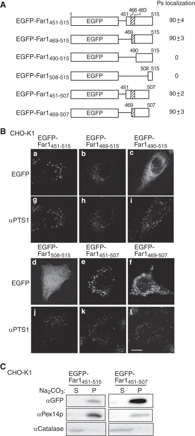
FIGURE 3.

The C-terminal portion of Far1 is sufficient for its peroxisome localization. A, schematic representation of the EGFP fusion proteins used to search for the minimum region of Far1 that is required for the localization of this protein to peroxisomes. The peroxisomal targeting activity of each fusion protein is represented by percentage. Data were collected by counting more than 100 cells expressing EGFP-fused Far1 mutants from three independent experiments and are represented by the mean ± S.D. Note that EGFP-Far1469–507 was localized to both peroxisomes and mitochondria. B, each construct was expressed in CHO-K1 cells for 14 h and detected by monitoring GFP fluorescence (a–f) and labeling with an anti-PTS1 antibody (g–l). Scale bar = 5 μm. C, the membrane integrities of two peroxisome-localizing fusion proteins expressed in CHO-K1 cells was assessed by carbonate extraction as described in Fig. 1B. P and S are membrane pellet and soluble fractions, respectively. Pex14p was used as a positive control for a peroxisomal integral membrane protein.
