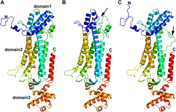
FIGURE 5.
Monomeric structure of wild type and models of ASL exon 2- or exon 7-deleted transcript variant. A, monomeric structure of WT ASL. ASL monomer has three distinct subdomains, domains 1 and 3 have similar structure and topology with two helix-turn-helix motifs in a perpendicular arrangement. Domain 2 has nine helices, and five of them form the central five-helix bundle with up-down-up-down-up topology. B, monomeric structural model of ex2del ASL. Two critical helices that are part of domain 1 (indicated by a black arrow) and contribute to the active site are missing in the ex2del variant of ASL. C, monomeric structural model of ex7del ASL. Exon 7 residues are part of domain 2 in the ASL monomer comprising the central five-helix bundle. Deletion of exon 7 results in a disordered central core with one of the five central helices partially replaced by an unstructured loop (indicated by a black arrow). N, N terminus; C, C terminus.

