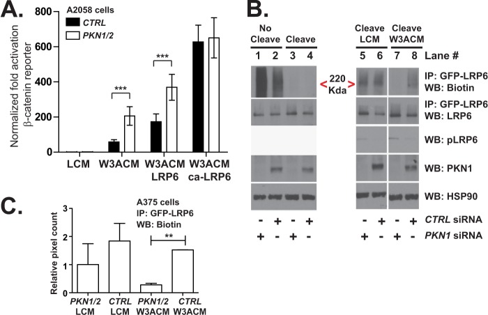
FIGURE 7.
PKN1 may attenuate Wnt/β-catenin signaling by inhibiting internalization of LRP6. A, 48 h following transfection of the siRNAs, A2058 cells were transiently transfected with plasmids expressing BAR, a constitutive promoter driving Renilla luciferase, and either empty vector, wild type LRP6, or truncated LRP6. The next day, luciferase abundance was assayed and plotted. The error bars represent S.D. from four replicates. B and C, HEK293T cells stably overexpressing GFP-LRP6 were transiently transfected with siRNA oligonucleotides targeting the indicated genes. 62 h later, the cells were serum-starved on ice and biotinylated with sulfo-NHS-SS-biotin. Following labeling, cells were stimulated and subsequently brought to 37 °C to initiate internalization of the biotinylated receptors. To cleave the remaining surface biotin, the samples were incubated with the reducing agent tris(2-carboxyethyl)phosphine. Lastly, cells were lysed, processed for immunoprecipitation (IP) using a GFP antibody, and analyzed by Western blot (WB). C, Western blots for biotin present in the GFP-LRP6 immunoprecipitation from two independent experiments were quantified by densitometry and plotted. Error bars represent S.E., and the p values (**, p < 0.01) were calculated using one-way ANOVA followed by Tukey's post test. In A, the p values (***, p < 0.001) were calculated using two-way ANOVA followed by a Bonferroni post-test. CTRL, control; W3ACM, WNT3A-conditioned medium; LCM, L cell-conditioned media; ca-LRP6, constitutively active LRP6.

