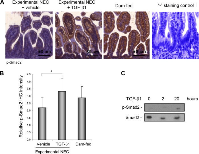
FIGURE 1.
Oral administration of TGF-β1 activated TGF-β signaling in IEC in experimental NEC. An animal model of NEC was conducted as described under “Materials and Methods.” Intestinal sections from experimental NEC pups treated with vehicle or 30 ng/ml TGF-β1 and from healthy dam-fed controls at the end of experiments on day 5 were IHC-stained for the active/phosphorylated form of the TGF-β intracellular mediator Smad2. A, representative staining shows increased p-Smad2 in IEC in experimental NEC pups gavaged with TGF-β1 as compared with the vehicle-treated control. Healthy dam-fed controls had basal activation of TGF-β signaling reflected by basal p-Smad2 levels. “−”, staining control of IHC was performed without using the primary antibody and shows the absence of background staining of p-Smad2. B, staining from A was quantified by scoring p-Smad2 on a 0–4 scale in three fields of each intestinal section (n = 3) as described under “Materials and Methods.” * depicts p < 0.05 by Student's t test. C, Smad2 was activated in IEC by 20 h following oral TGF-β1 administration. Intestine was collected at 0, 2, and 20 h after the first gavage of TGF-β1. IEC were isolated from intestine as described under “Materials and Methods,” lysed, and subjected to immunoblotting for total and phospho-Smad2.

