
FIGURE 7.
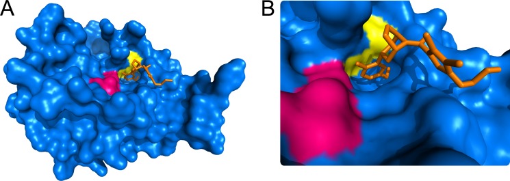
A, surface rendering of the βII-tryptase monomer showing the active site residues in magenta, the Cys220–Cys248 disulfide bond in yellow, and an inhibitor bound in the active site in orange (Protein Data Bank identifier 3V7T) (62). B, close-up of the active site. The structure was generated using PyMOL v0.99 (DeLano Scientific LLC).