FIGURE 6.

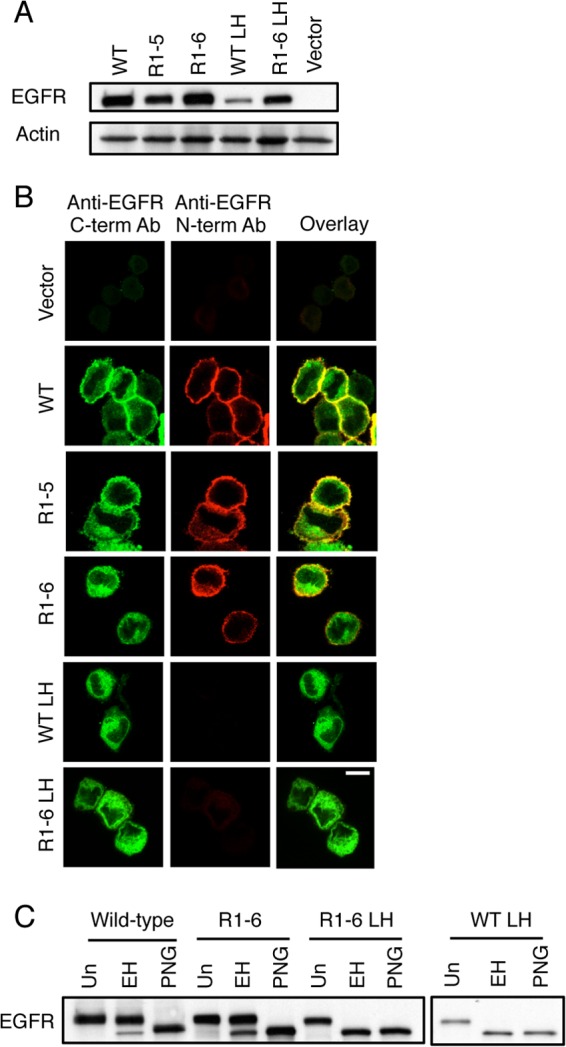
EGFR Mut R1-6/L393H is retained in the ER when stably expressed in NIH 3T3 cells. A, whole cell lysates of NIH 3T3 cells stably expressing WT EGFR, EGFR Mut R1-5, EGFR Mut R1-6, EGFR L393H (LH), EGFR Mut R1-6/L393H, or vector only were subjected to Western blot analysis using an anti-EGFR antibody and an anti-actin antibody (loading control). B, confocal images of NIH 3T3 cells stably expressing vector only, WT EGFR, EGFR R1-5, EGFR Mut R1-6, EGFR L393H, or EGFR Mut R1-6/L393H. Before imaging cells were fixed and labeled with an anti-N-terminal EGFR antibody followed by Alexa 647 anti-mouse IgG, then washed, permeabilized, and labeled with anti-C-terminal EGFR antibody followed by Alexa 488-anti-rabbit IgG. Scale bar, 10 μm. C, whole cell lysates from NIH 3T3 cells stably expressing either the WT EGFR, EGFR Mut R1-6, EGFR Mut R1-6/L393H, or EGFR L393H were untreated (Un) or were treated with Endo H (EH) or with PNGase F (PNG) for 24 h before Western blot analysis with an anti-EGFR antibody.
