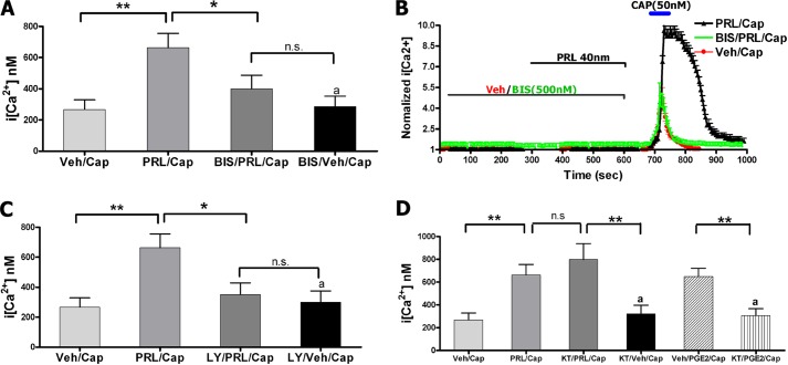
FIGURE 1.
A, intracellular calcium ([Ca2+]i) accumulation by capsaicin (Cap; 50 nm) in TG neurons from female rats pretreated for 5 min with vehicle (Veh), PRL (40 nm (1 μg/ml); BIS (0. 5 μm), or PRL+BIS. CAP was applied for 1 min. Statistical analysis was performed using a one-way ANOVA with Bonferroni's post hoc test (*, p < 0.05; **, p < 0.01; n.s., nonsignificant; n = 40–60). Error bars, S.E. B, representative Ca2+ imaging traces from TG neurons responsive to CAP after pretreatment for 5 min with vehicle, PRL, or PRL+BIS. Durations of CAP and PRL applications and concentrations are indicated. C, CAP-evoked [Ca2+]i rise in TG neurons pretreated for 5 min with vehicle, PRL, LY294002 (LY; 20 μm), or PRL+LY. CAP was applied for 1 min. One-way ANOVA with Bonferroni's post hoc test was used (*, p < 0.05; **, p < 0.01; n.s., nonsignificant; n = 40–60). D, CAP-evoked [Ca2+]i rise in TG neurons pretreated for 5 min with vehicle, PRL, KT-5720 (KT; 0.2 μm), or PRL+KT. The positive control was regulation of CAP-evoked [Ca2+]i rise by prostaglandin E2 (PGE2; 1 μm), and blockade of this effect by KT. CAP was applied for 1 min. One-way ANOVA with Bonferroni's post hoc test was used (**,p < 0.01; n.s., nonsignificant; n = 30–60).

