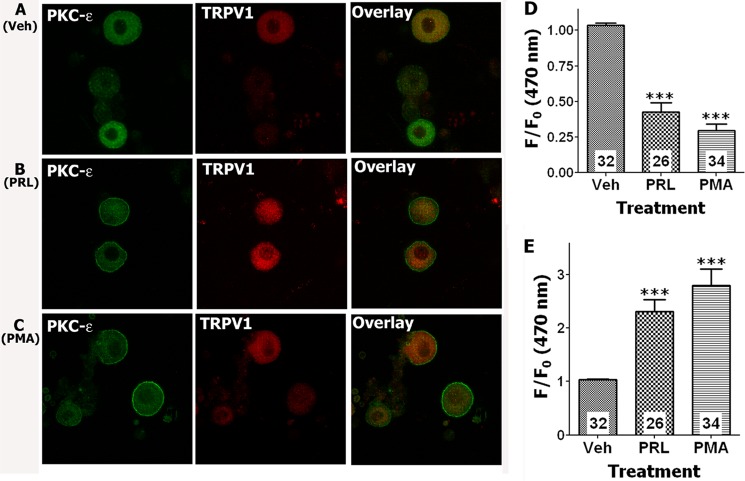
FIGURE 5.
A–C, PRL evokes the translocation of PKCϵ to the plasma membrane in cultured TRPV1-positive TG neurons from female rats. Confocal image of TG neurons was taken at 120× magnification. TG neurons were treated for 5 min at 37 °C with vehicle (Veh), PRL (1 μg/ml), or PMA (0.5 μm). Following treatments, TG neurons were fixed with 4% formalin and processed for IHC with antibodies against PKCϵ and TRPV1. Treatment with vehicle produced no translocation of PKCϵ (A). PRL (B) and PMA (C) induced translocation of PKCϵ to the plasma membrane in a subset of TRPV1-positive TG neurons. D and E, quantification of translocation (f/f0 at 470 nm) for PKCϵ is presented for cytoplasmic (D) and plasma membrane regions (E). Number of analyzed cells are indicated within bars. One-way ANOVA with Bonferroni's post hoc test was used (***, p < 0.001). Error bars, S.E.

