Fig. 5.
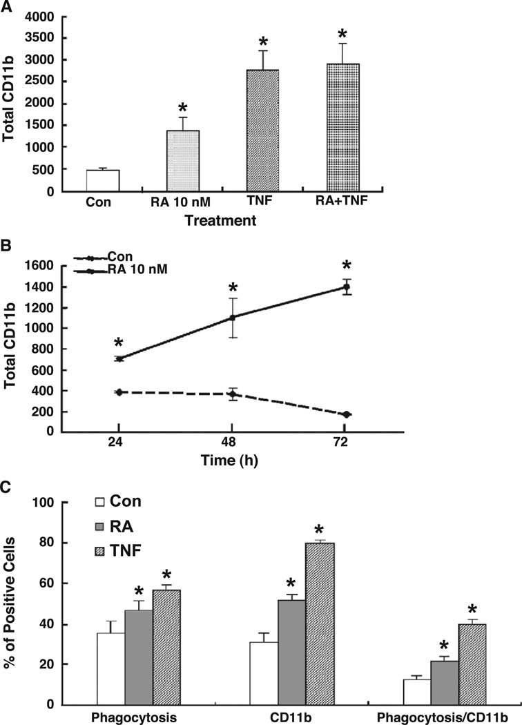
CD11b expression level and phagocytosis activity in retinoic acid-treated THP-1 cells. THP-1 cells were plated in low serum condition and then treated with and without RA (10 nM) in the present of 10% FBS. (A) CD11b expression levels in THP-1 cells. Cells treated with and without RA for 48 h were subjected to flow cytometry after staining with an anti-CD11b antibody. The total expression level was calculated by multiplying the percentage of CD11b-positive cells by the average fluorescence intensity per cell. (B) CD11b level in THP-1 cells after treated for 24 to 72 h. (C) Phagocytic activity of RA-treated THP-1 cells. THP-1 cells were treated with or without RA for the times indicated before fluorescein-labeled E. coli bioparticles were added for an additional 2 h. After harvesting, the cells were subjected to CD11b staining and then analyzed by flow cytometry. The values shown are the mean ± SD of three independent experiments performed in triplicate. * P < 0.05.

