Fig. 3.
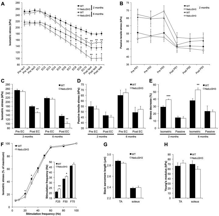
Biomechanical studies of skeletal muscle function in WT and NebΔSH3 mice. (A) Time course of isometric stress measured before (Pre Iso1–3), during (EC1–10) and after (Post Iso1–3) cyclic eccentric exercise of the fifth toe EDL muscle from 2- and 6-month-old WT and NebΔSH3 mice. Each symbol represents the mean ± s.e.m. of six mice per group. (B) Passive tensile stress (PS) at 15% passive stretch before (Pre PS1–3) and after (Post PS1–3) eccentric exercise. (C) Average maximum isometric stress production before and after eccentric exercise. (D) Average passive tensile stress at 15% stretch before and after eccentric exercise. (E) Magnitude of eccentric contraction-induced injury, defined as the percentage decrease in isometric stress and/or passive tensile stress after eccentric exercise. (F) Force–frequency relationship in the fifth toe EDL muscle from 2-month-old WT and NebΔSH3 mice. Inset: NebΔSH3 muscle requires higher stimulation frequency to achieve 25% and 50% of maximum isometric stress production (F25, F50). n = 6 (A–F). (G,H) Passive mechanical properties of single fibers from TA and soleus muscle from 2-month-old WT (n = 9) and NebΔSH3 (n = 9) mice. No significant differences in slack sarcomere length (G) or Young's modulus (H) between WT and NebΔSH3 fibers. *P<0.05, **P<0.01, ***P<0.001.

