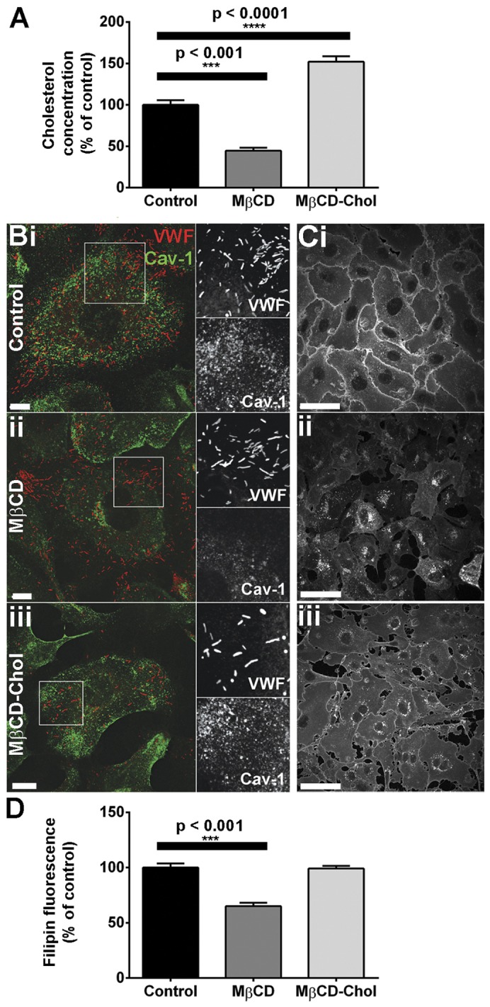
Fig. 5.

Depletion or supplementation of HUVEC cholesterol by MβCD. (A) HUVECs were treated with vehicle (control), 5 mM MβCD or 5 mM MβCD-Chol for 30 minutes and cholesterol content was quantified as described in the Materials and Methods. Data were compared by one-way ANOVA (n = 7 experiments for control, n = 3 for MβCD and n = 12 for MβCD-Chol). (B,C) HUVECs treated as described for A were stained with specific antibodies to VWF (red, sheep Ab) and Cav-1 (green) (B) or filipin (C). (D) Filipin fluorescence was quantified for each treatment condition and was compared by one-way ANOVA (n = 75 cells for control, 80 for MβCD and 120 for MβCD-Chol). Scale bars: 10 µm (B), 50 µm (C).
