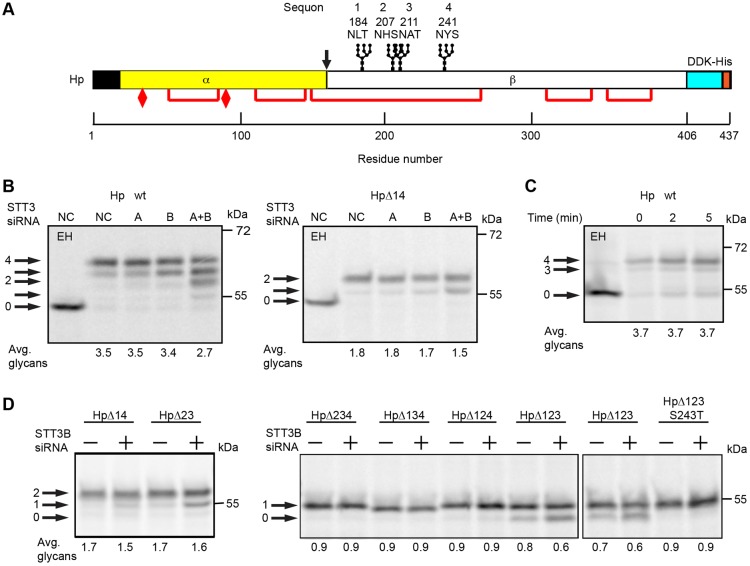
Fig. 4.
Glycosylation of haptoglobin. (A) Diagram of the haptoglobin precursor (Hp) showing the signal sequence (black), protease processing site at the α-β junction (arrow), glycosylation sites, disulfide bonds (red lines), cysteine residues that form interchain disulfides to link two α-subunits (elongated diamonds) and a DDK-His tag. The four sequons are numbered 1–4; Hp mutants lacking one or more sequons are designated as HpΔXYZ, where XYZ is the list of mutated sequon(s). (B,D) HeLa cells were treated with negative control (NC) siRNA or siRNAs specific for STT3A or STT3B as indicated for 48 hours prior to transfection with Hp expression constructs. Cells were then pulsed for 4 minutes and chased for 20 minutes. (C) Cells expressing Hp-wt were pulse labeled for 4 minutes and chased as indicated. Hp glycoforms were precipitated with anti-DDK sera and resolved by SDS-PAGE. EH designates digestion with endoglycosidase H. Quantified values below gel lanes are for the displayed image that is representative of two or more experiments.

