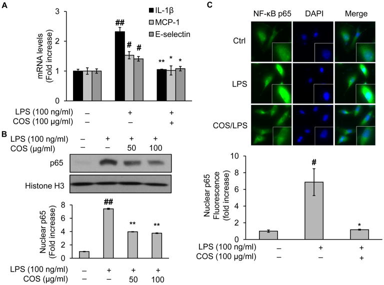
Fig. 1.
COS inhibit NF-κB nucleus translocation and inflammatory cytokine gene expression in LPS-treated endothelial cells. EA.hy926 or BAEC were pretreated with COS (up to 100 μg/ml) for 4 h followed by LPS (100 ng/ml) incubation for 4 h. Then the cells were subjected to the assessment of (A) mRNA levels of inflammatory cytokines including IL-1β, MCP-1, and E-selectin, using RT-PCR as described in detail in Materials and Methods (Inflammatory cytokine gene expression analysis) in EA.hy926, and in BAEC: (B) Western blotting of NF-κB in the nucleus fraction prepared by NE-PER® Nuclear and Cytoplasmic Extraction Reagents, and (C) immunofluorescent staining of NF-κB or DAPI with a kit including ProLong® Gold and SlowFade® Gold Antifade. The secondary antibody was conjugated with a fluorescent green dye (Alexa Fluor 488). Nuclear p65 fluorescence was quantified. All images shown are representative of three independent experiments. Data are expressed as means ± SEM. ##P<0.01 compared to the vehicle-treated control group; *P<0.05, **P<0.01 compared to the LPS-only group.

