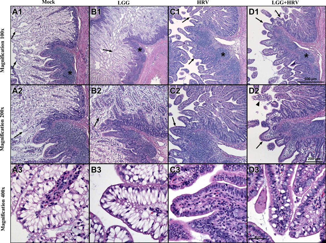
Fig. 2. Histopathology of ileum of Gn pigs on PID 2.
Series of photomicrographs of mucosa of ileum from a mock inoculated control (A), LGG alone (B), HRV alone (C) and LGG+HRV (D) exposed Gn pigs euthanized on PID 2 were acquired from H& stained cross-sections. The sections include a region containing Peyer's patch (* A1, B1, C1, D1) and adjacent mucosa. Note the prominence of pale-stained vacuolated enterocytes lining long, thin villi in the mock exposed control and LGG-exposed animals (arrows A1–2, B1–2 and shown at higher magnification A3, B3). In the two HRV infected groups the vacuolated cells are largely lost, and villi are shortened, blunted and have increased lymphocytes in the lamina propria (arrows in C1–2, D1–2). There is modest preservation of vacuolated enterocytes in LGG+HRV pigs (arrowhead in D2 and higher magnification image in D3) compared to HRV alone pigs (C2–3). Scale bar for each row of images is seen in D1–3.

