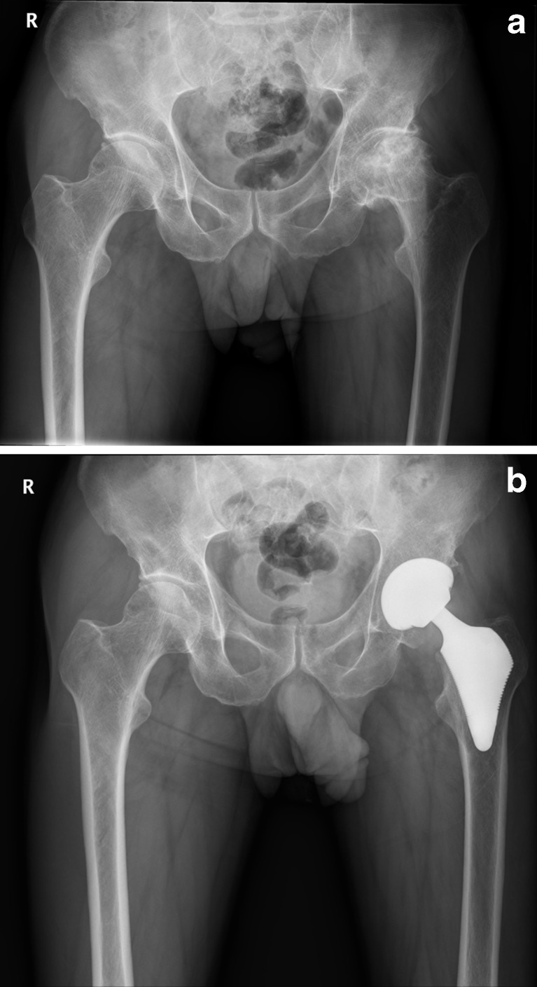
Fig. 2.
Radiographs of 75-year-old man with osteonecrosis of left femoral head. a Preoperative radiograph of left hip demonstrates collapse of left femoral head (Ficat stage IV) and Dorr type C bone. b Postoperative radiograph of left hip obtained nine years after surgery demonstrates that Proxima stem is rigidly fixed in a satisfactory position in left hip

