
Fig. 4.
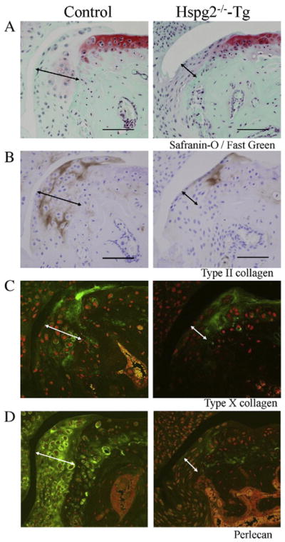

Perlecan deficiency reduces chondrocyte differentiation, cell proliferation, and Smad2 phosphorylation during osteophyte development. Histological sections of the knee joints of control and Hspg2−/−-Tg mice 4 weeks after surgery (OA). Safranin-O staining (red) with fast green (green) counter staining (A), immunohistochemistry for type II collagen (COLII) (B), type X collagen (COLX) (C), perlecan (D), PCNA (E), and phosphorylated Smad2 (p-Smad2) (F). COLII (brown, B) and COLX (green, C) expression in osteophytes was reduced in Hspg2−/−-Tg mice compared with control mice. Perlecan expression (green, D) was detected in synovial cells and chondrocytes of osteophytes with higher expression levels in hypertrophic chondrocytes of control mice. In Hspg2−/−-Tg mice, perlecan expression was absent in osteophytes but was detected in COLII-expressing chondrocytes (B and D) because these cells express the Hspg2-transgene driven by the Col2a1 promoter/enhancer. Fewer PCNA-positive cells (brown) were observed in osteophytes of Hspg2−/−-Tg mice compared with control mice (E). Quantitative analysis showed that the number of PCNA-positive cells in osteophytes was significantly reduced in Hspg2−/−-Tg mice compared with control mice (G). P-Smad2 staining (brown) was reduced in Hspg2−/−-Tg mice compared with control mice (F). Quantitative analysis confirmed the significant reduction of p-Smad2 in osteophytes of Hspg2−/−-Tg mice (H). The osteophytes are shown with black lines (A, B, E and F) and white lines (C and D). Scale bars, 100 μm. All data represent means and SEM.