
Fig. 5.
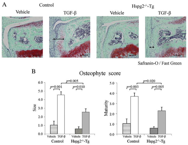
Reduction in TGF-β-induced osteophyte formation in Hspg2−/−-Tg mice. Histological sections of the knee joint 1 week after injection with either TGF-β or vehicle in control and Hspg2−/−-Tg mice (A). Safranin-O staining and osteophyte size and maturity were reduced in the joints of Hspg2−/−-Tg mice compared to control mice (A). Quantification showed that osteophyte size and maturity on the TGF-β injection side (TGF-β) of Hspg2−/−-Tg mice were significantly reduced (B). The osteophytes are shown with black lines (A). Scale bar, 100 μm. All data represent means and SEM.