
Figure 1.
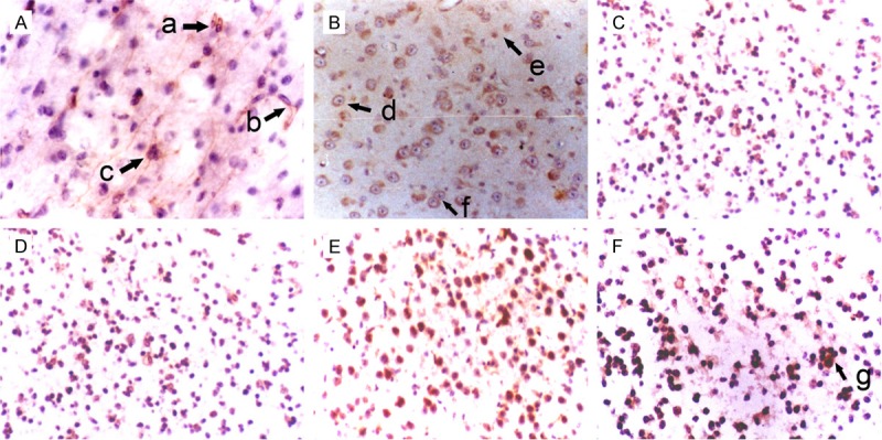
Morphology of NSCs in the hippocampus of fetuses with different gestational age (SABC, DAB staining, ×200). A: 36 weeks; B: 32 weeks; C: 28 weeks; D: 24 weeks; E: 20 weeks; F: 16 weeks. a: Oval NSCs; b: NSCs interacted with each other via synapses; c: Colony formation for 3 NSCs; d: Big and round NSCs; e: Small and round NSCs; f: Symmetrical division of NSCs; g: Colony formation from several NSCs.