
Figure 1.
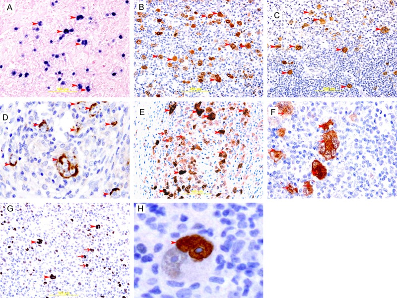
Expression of EBV, COX2, p16 and p53 in cHL. A: Representative section of cHL showing blue nuclear staining of H/RS cells (arrow head) for EBER, In situ hybridization method. B: Representative section of cHL showing brown membranous and cytoplasmic staining of H/RS cell (arrow head) for LMP-1, Immunoperoxidase streptavidin-biotin method. C&D: Representative section of cHL showing brown cytoplasmic staining of H/RS cell (arrow head) for COX2, Immunoperoxidase streptavidin-biotin method. E&F: Representative section of cHL showing brown nuclear (arrow head) and cytoplasmic (thin arrow) staining of H/RS cell for p16, Immunoperoxidase streptavidin-biotin method. G: Representative section of cHL showing brown nuclear staining of binucleated RS cells (arrow head) and mononucleated H/RS cells (thin arrows ) for p53, Immunoperoxidase streptavidin-biotin method. H: A binucleated RS cell showing strong nuclear staining (arrow head) for p53 in one nucleus, please note, big prominent nucleolus is not stained for p53.