
Figure 5.
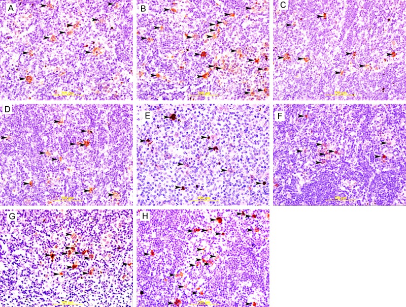
Cleaved caspase 3 staining in cHL. A&B: Showing cytoplasmic immunoreactivity of H/RS cells (arrow head) to cleaved-caspase in EBV+ve and EBV-ve HL respectively. There is significant increase in cleaved-caspase-positive H/RS cells (arrow head) in EBV-ve HL. Streptavidin-biotin immunoperoxidase method. C&D: Showing cytoplasmic immunoreactivity of H/RS cells (arrow head) to cleaved-caspase in COX2+ve and COX2-ve HL respectively. There is no significant difference in cleaved-caspase-positive H/RS cells (arrow head) between COX2+ve and COX2-ve HL. Streptavidin-biotin immunoperoxidase method. E&F: Showing cytoplasmic immunoreactivity of H/RS cells (arrow head) to cleaved-caspase in p16+ve and p16-ve HL respectively. There is no significant difference in cleaved-caspase-positive H/RS cells (arrow head) between p16+ve and p16-ve HL. Streptavidin-biotin immunoperoxidase method. G&H: Showing cytoplasmic immunoreactivity of H/RS cells (arrow head) to cleaved-caspase in p53+ve and p53-ve HL respectively. There is significant increase in cleaved-caspase-positive H/RS cells (arrow head) in p53-ve HL. Streptavidin-biotin immunoperoxidase method.